Case wise Active learning and discussion-2
Case 2-- Paraparesis
23 yr old with bilateral lower limbs weakness associated with tingling and numbness
We the MBBS final year students have been given these cases on weekly basis to solve in an attempt to understand the topic of "patient clinical data analysis" to develop my competency in reading and comprehending clinical data including history, clinical findings, investigations and come up with a diagnosis and treatment plan.
- Link to Reference:
- Link to my analysis:
ACTIVE LEARNING AND CONVERSATIONAL DECISION SUPPORT TO TREATING TEAM OF THIS CASE :
- My active learning discussion:
[5/22, 23:51] MBBS 2016 UG 3: Sir was the lumbar puncture done in the patient to diagnose TB?
[5/22, 23:53] Post residency PG1: Check out the conversations with your colleagues posted here
https://medicinedepartment.blogspot.com/2020/05/frequently-asked-questions-around-case.html?m=1 and you will get the answer
[5/22, 23:58]MBBS 2016 UG 3: Ok sir...thank you
- OTHER DISCUSSIONS:
[5/23, 10:13 AM] PG Post Residency 1 : First let me share some discussion with another student below
"Good morning sir!
Sir, why was there no biopsy taken from the abscess sites? To check for what bacteria might've caused it?"
" Because I'm not able to visualize being able diagnose tuberculosis with the given data."
" Check out the MRI report"
" Sir. Isn't there also a possibility that during the I&D for gluteal abscess there was damage to L4 and 5 which lead to the paraparesis?"
" Very Good question. Ask our interns and PGs. Tell me if you need their numbers"
" Yes sir. Because in a few cases I've read
And most of them also had no AFB"
" How sensitive and specific is that and would it be useful for our patient?"
" Sir. The pus from the pyocele was sent for culture sensitivity and revealed no AFB it seems sir"
" Tuberculous abscesses are only diagnosed on the basis of histo pathology"
" Okay sir. I shall ask her"
" The question is why didn't we send a biopsy of the abscess tract to get a better histopathology diagnosis"
"I have the asked for the report from the intern, and she is yet to reply."
" They also had positive tuberculin Skin test sir"
" It's already there in one of their e logs shared with you"
[5/23, 10:17 AM] mbbs 2016 UG1 : It's not very sensitive because most of us have received a BCG vaccine, which would give us a false positive
[5/23, 10:18 AM] mbbs 2016 UG1: Did we assume he has TB? There seems to be no substantial evidence to prove it.
[5/23, 10:18 AM] PG post residency 1 : Can you share some literature that says BCG vaccination gives false positive Mantoux test.
Also is being Mantoux or tuberculin positive the same as being diseased?
[5/23, 10:30 AM] mbbs 2016 UG1: I found two conflicting studies :
https://pubmed.ncbi.nlm.nih.gov/28087302/?from_single_result=28087302&expanded_search_query=28087302
The above study provides evidence that BCG vaccination after infancy may influence TST results beyond the 10-year period conventionally accepted by the Centers for Disease Control and Prevention (CDC), extending up to 55 years after vaccination. This suggests that BCG vaccination should be taken into account when interpreting TST results regardless of the time elapsed since vaccination.
https://pubmed.ncbi.nlm.nih.gov/17131776/?from_single_result=17131776&expanded_search_query=17131776
Whereas, this study claims that The effect on TST of BCG received in infancy is minimal, especially > or =10 years after vaccination. BCG received after infancy produces more frequent, more persistent and larger TST reactions.
[5/23, 4:14 PM] PG Post Residency 1: 👍👏👏
Ok now let's back to solving the problem of confirming the diagnosis of tuberculosis in the patient. What would have been the best way forward to prove tuberculosis in this patient instead of beginning the treatment empirically?
[5/23, 4:23 PM] mbbs 2016 ug1: CBNAAT
[5/23, 4:28 PM] PG Post residency 1: CBNAAT of what?
[5/23, 4:33 PM] mbbs 2016 ug1: of sputum
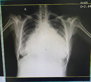
[5/23, 4:35 PM] mbbs 2016 ug1: Sputum smear for AFB and Chest X-ray should also be performed
[5/23, 4:50 PM] PG Post residency 1: Wasn't it performed? Please ask the interns and PGs who made the log
[5/23, 4:54 PM] mbbs 2016 ug1: Alright sir.
[5/23, 4:57 PM] mbbs 2016 ug1: And sir, how is this related to Vasculitis?
[5/23, 4:59 PM] PG POST RESIDENCY 1:
Conversation with another student "So is it ACA Vasculitis as mentioned by one of my classmates??"
[5/23, 4:59 PM] PG POST RESIDENCY 1: "I mean is the diagnosis Tubercular ACA vasculitis in the medial Homenculus area of cerebral cortex??"
[5/23, 4:59 PM] PG POST RESIDENCY 1: "https://www.ncbi.nlm.nih.gov/pmc/articles/PMC4606267/
This link she mentioned also tells about cerebral vasculitis without TBM"
[5/23, 4:59 PM] PG POST RESIDENCY 1: "Yes share the interesting learning points that you gained from that article 👍"
[5/23, 5:01 PM] mbbs 2016 ug1 Thanks Sir!
[5/23, 5:30 PM] mbbs 2016 ug1: Here are a few doubts I have,
[5/23, 5:40 PM] mbbs 2016 ug1: According to the MR images shared, there seems to be no evidence that the region of the cerebral cortex supplied by the ACA is affected to conclude that it is the cause of the paraparesis.
[5/23, 6:24 PM] KIMs 2016 ug1: The Anatomical location of the UMN lesion could be anywhere - Primary Motor Cortex, Internal Capsule, Midbrain, Pons , Lateral Corticospinal Tract
[5/23, 6:29 PM] KIMs 2016 ug1: The pathology could be -
1. Damage to the brain - due to Thrombus, Hemorrhage (Stroke), Infection, Tumour (Cancer), PML
2. Injury to the brainstem or white matter of the spinal cord - due to Multiple Sclerosis, Transverse Myelitis, Trauma, Spinal Stenosis, Spinal Abscess
[5/23, 6:36 PM] KIMs 2016 ug1: How do we localise the anatomical location affected in this UMN lesion which may have caused the paraparesis? Is the only was to do so by performing an MRI of the brain and the spinal cord? How does one interpret it?
[5/23, 6:43 PM] KIMs 2016 ug1: What is the relationship between the psoas absess and "the ring enhancing lesions seen in the right and left cerebral hemispheres" on MRI?
[5/23, 7:02 PM] KIMs 2016 ug1: Mostly, The vertebral lesion could not be producing any neurological signs because according to the MRI spine report , the patient has L4, L5 Spondylodiscitis which means, most likely, the spinal cord could not have been involved as it ends at the level of L1,L2 vertebrae. Though, the cause of the Psoas Abscess and Gluteal Abscess (Both cold abscesses) can be explained by spread of AFB from the L4,L5 Spondylodiscitis as commonly seen in Tuberculous spondylodiscitis.
[5/23, 7:54 PM] pg post residency 1: 👍Yes the last paragraph is spot on
[5/23, 7:54 PM] pg post residency : The ring enhancing lesions could be tuberculoma and the psoas abscess is also due to mycobacteria
[5/23, 7:55 PM] pg post residency 1: Have you checked the report? The very few films that have been shared may not reveal all
[5/23, 7:59 PM] pg post residency 1: Yes but the first step is to localize it anatomically and the clinical possibilities were bilateral leg area of the brain and high cervical cord. Brain stem was unlikely as there was no cranial nerve involvement.
Once the localization is over then the pathology can range from vascular, demyelinating, degenerative to neoplasia
[5/23, 8:17 PM] KIMs 2016 ug1: I've only seen the images shared on the blog by Vaishnavi ma'am (intern)
[5/23, 8:21 PM] KIMs 2016 ug1: What symptoms or signs may the Tuberculoma be causing?
[5/23, 8:24 PM] pg post residency: Check the report. Or ask Vaishnavi to share the radspa link to the entire mri images with you
[5/23, 8:25 PM] pg post residency1: Good question. Currently none that we know of
[5/23, 8:33 PM] KIMs 2016 ug1: In the Brain, that would be the Precentral Gyrus in the frontal lobe. What points us towards a high cervical cord lesion?
[5/23, 8:34 PM] KIMs 2016 ug1: One possibility could be Vomiting (which was described as non-projectile by the patient but could be projectile in actuality)
[5/23, 8:38 PM] pg post residency 1: UMN signs that are above the C5 cervical cord as biceps C5 is exaggerated.
Above the high cervical cord comes the brain stem which is ruled out as there are no cranial nerve involvement and finally bilateral cortical leg area is the only location left
[5/23, 8:38 PM] pg postresidency 1: Why would the ring enhancing lesions cause vomiting?
[5/23, 8:45 PM] KIMs 2016 ug1: Due to raised ICT
[5/23, 8:47 PM] pg postresidency 1: Do ring enhancing lesions cause raised ICT without any focal neurological deficit?
[5/23, 8:47 PM] KIMs 2016 ug1: Vasculitis of the ACA explains the lesion at the bilateral cortical leg area.
But, what could be the cause for a lesion above the C5 cervical cord?
[5/23, 8:51 PM] pg postresidency 1: When someone has quadriparesis due to an apparent UMN lesion above C5 then the differentials become either high cord lesion or brain stem lesion or cortical or internal capsular lesion
[5/23, 8:51 PM] KIMs 2016 ug1: No probably not. But I don't know why.
[5/23, 9:45 PM] KIMs 2016 ug1: I still don't understand how he was diagnosed with TB, sir.
[5/23, 10:12 PM] KIMs 2016 ug1: I asked Rashmitha ma'am whether Sputum Smear for AFB and CBNAAT was done and she said that the sputum was not inducible, so it wasn't possible and that the abscess pus was negative.
[5/23, 10:17 PM] pg postresidency 1: 👍
What other disease can produce such pus everywhere and also spread to the brain?
[5/23, 10:19 PM] KIMs 2016 ug1: What about Sarcoidosis?
[5/23, 10:21 PM] Rakesh Biswas: Share a similar case reported with sarcoidosis
[5/24, 3:09 PM] KIMs 2016 ug1: https://www.ncbi.nlm.nih.gov/pmc/articles/PMC3669974/
[5/24, 3:09 PM] KIMs 2016 ug1: dDx : Spinal Sarcoidosis - Osseous spinal sarcoidosis: an unusual but important entity to remember
https://www.ncbi.nlm.nih.gov/pmc/articles/PMC3030116/
[5/24, 3:15 PM] KIMs 2016 ug1: Other questions I had are:
1. What pathology could be responsible for a lesion at or above the C5 cervical cord? Vascular, demyelinating, degenerative, neoplasia? Was anything visible on radiology?
2. How do we rule out various pathologies after we have located the anatomical location of the lesion causing the UMN symptoms (paraparesis)? Which in this case is the bilateral leg area of the brain.
3. Why didn't we do a biopsy for histopathological diagnosis of the affected vertebrae showing spondylodiscitis (L4,L5) on MRI?
Because as I mentioned, any pathogenic organism could have caused it.
4. Why don't ring enhancing lesions cause raised ICT without any neurological deficit?
[5/24, 3:39 PM] pg post residency 1: Did this patient in the report have spinal cord or peripheral nerve involvement due to sarcoid?
[5/24, 3:50 PM] pg post residency 1 : Very good questions.
1) For that particular patient no pathology was visible in any part of the spinal cord although clinically one strong differential was high cervical cord due to the clinical neurological findings of localization.
2. Ultimate diagnosis was always made in the past by biopsy either through autopsy in case of unfortunate death of the patient or through operative biopsy. Because getting those are invasive and difficult the other option in medical decision making is to go by statistical probability of the pathology.
3) As stated above because of high statistical probability of a treatable infectious pathology known to cause similar findings in the past, most physicians avoid invasiveness and favor tolerating moderate diagnostic uncertainty over the ability to provide potential relief through easily available standard course of treatment.
4) If those ring enhancing lesions were tumors or inflammed infective lesions such as neurocysticercosis or tuberculoma (ours was possibly a non inflammed tuberculoma) they can cause raised ICT. Focal deficits are seen in ring enhancing lesions when they impinge on the pyramidal pathway. I guess our previous discussion around this was incomplete and hence the confusion. Thanks for diligently pursuing it. 👍"
Mbbs 2016 UG student 3:
2) [5/20, 8:55 AM] mbbs 2016 UG student 3: Sir in this case
https://vaish7.blogspot.com/2020/05/medicine.html?m=1, the patient didn’t have fever or any symptoms of inflammation how would we think of TB?
[5/20, 9:17 AM]: MD PG (post residency) That's the idea of differential diagnosis.
One discusses the differentials to capture something the current symptoms wouldn't allow us to think of from what is currently known.
[5/20, 8:54 AM] +91 0: https://vaish7.blogspot.com/2020/05/medicine.html?m=1
[5/20, 11:03 AM] +91 : Why haven’t we done a Lumbar puncture in the above patient sir?
[5/20, 4:40 PM] : MD PG (post residency
Alright. How do you think it would have helped if we did the lumber puncture? What were you expecting to find in it?
[5/20, 4:40 PM] : Lymphocyte?
To confirm TB
[5/20, 4:59 PM] : MD PG (post residency
How many are normal in CSF?
How does TB lead to lymphocytosis in CSF?
[5/20, 5:04 PM] : Less than 5 per ml is normal in CSF
Meningeal inflammation sir.
[5/20, 5:07 PM] MD PG (post residency:
So are you suspecting meningitis here? Does this patient of paraparesis have any sign of meningitis? What signs would you look for?
[5/20, 5:49 PM] mbbs 2016 UG: Kernigs sign
[5/20, 6:26 PM] MD PG (post residency
: So does this patient have any signs and symptoms of meningitis to merit an LP. You can even ask the intern. I can share her number if you want. You can message her or better is ask her in her log book comment box
[5/20, 7:16 PM] : No sir.
[5/20, 7:16 PM] : Yes sir I’ll ask her.
[5/20, 8:05 PM] MD PG (post residency
: 👍keep your questions coming. These are important to score learning points
[5/20, 8:55 PM] +91: Sir is it falx cerebri tuberculoma?
[5/20, 8:57 PM] MD PG (post residency
: Are there descriptions of such a location being affected by mycobacteria in the past on your review of literature?
[5/20, 8:57 PM]: Yes sir
[5/20, 8:57 PM]: It says there are 3 cases reported as now.
[5/20, 8:59 PM] MD PG (post residency:
Share those cases and check out the similarities with our patient
[5/20, 9:03 PM] mbbs 2016 UG: The patient had monoperesis because it was unilateral.
[5/20, 9:03 PM] MD PG (post residency
: Tell me the similarities and dissimilarities and always share the links to articles as we can't share the PDFs in your log book comment box or the FAQ learning points page
[5/20, 9:04 PM]: mbbs 2016 UG: http://www.roneurosurgery.eu/atdoc/CucuA_Falx.pdf
[5/20, 9:06 PM] mbbs 2016 UG 3: Sir he had monoplegia initially
Later developed seizure which our patient didn’t.
CT revealed edema and a tumor like mass near the falx cerebri.
[5/20, 9:08 PM] MD PG (post residency
: Was the second line similar to our current patient?
[5/20, 9:17 PM] Yes sir
[5/20, 9:17 PM] He had meningeal enhancement and edema.
[5/20, 10:51 PM] MD PG (post residency)
: Yes but is that same as having a tumor like mass near the falx?
[5/20, 11:11 PM] mbbs 2016 UG3: Enhancing lesion is tumor like?
[5/20, 11:13 PM] MD PG (post residency)
: Not as seen in the mri of our patient? Check out how a tumor enhancement looks like
[5/20, 11:15 PM] mbbs 2016 UG 3: There’s a diffuse enhancement in our patient
[5/20, 11:22 PM] Rakesh Biswas: Is that what happens in tumors?
[5/20, 11:22 PM] Kims 2016: No sir.
[5/20, 11:23 PM] Rakesh Biswas: Was contrast enhancement demonstrated in our patient's mri study? Was our patient given contrast?
[5/20, 11:24 PM] Kims 2016: The blog says there was significant enhancement
[5/20, 11:27 PM] Rakesh Biswas: Please clarify this with the intern who wrote that web log
[5/21, 8:18 AM] Kims 2016: Okay sir
[5/21, 8:49 AM] Kims 2016: Sir another doubt,
When we did an Xray abdomen and it showed psoas abscess on the right why didn’t we assume it to be the reason for the lower limb weakness and did an MRI?
[5/21, 8:54 AM] Rakesh Biswas: They happened at the same time but tell me what are your thoughts on the anatomical location of his lesion that is actually causing his paraparesis?
[5/21, 8:56 AM] Kims 2016: Psoas abscess compressing the nerves of the cord.
[5/21, 8:57 AM] Kims 2016 : It can also be because of the ring enhancing lesions of the cerebrum.
[5/21, 8:57 AM] Rakesh Biswas: Which part of the cord? Where is the psoas located and how does it compress the cord?
[5/21, 8:58 AM] Kims 2016: Not the cord but the nerves arising from the cord.
L4 and L5
[5/21, 8:58 AM] Rakesh Biswas: Which is the exact location? You can't say the paraparesis is both UMN as well as LMN?
[5/21, 8:59 AM] Kims 2016: Parasaggital?
[5/21, 9:01 AM] Kims 2016: It can be both.
[5/21, 9:03 AM] Kims 2016: In ALS, Neurodegenration, Spastic paraplegia
[5/21, 9:08 AM] Rakesh Biswas: What clinical signs does he have? Does he have both UMN and LMN signs?
[5/21, 9:09 AM] Kims 2016: UMN - Hypotonia, Absent reflexes
[5/21, 9:10 AM] Rakesh Biswas: 😳
[5/21, 9:12 AM] Kims 2016: LMN sir😬 sorryy
"[5/21, 9:13 AM] Rakesh Biswas: He has hypotonia and absent reflexes? 😳🤔
[5/21, 9:13 AM] Kims 2016 : Ankle clonus was absent
[5/21, 9:15 AM] Kims 2016 : And in the examination she wrote hypotonia in lower limbs.
[5/21, 9:42 AM] Rakesh Biswas: Which patient are you talking about? This one with psoas abscess? 🤔
Can you share the link to the case you are discussing?
[5/21, 9:42 AM] Kims 2016 : https://vaish7.blogspot.com/2020/05/medicine.html?m=1
[5/21, 9:42 AM] Kims 2016: This one sir.
[5/21, 9:49 AM] Rakesh Biswas: Yes but what about the reflexes?
[5/21, 9:50 AM] Kims 2016: Reflexes were present sir.
[5/21, 9:51 AM] Rakesh Biswas: Then can it be LMN?
[5/21, 9:51 AM] Kims 2016: No sir
But the reflexes were not exaggerated also
And there was hypotonia.
[5/21, 9:54 AM] Rakesh Biswas: Can a paraparesis be due to LMN as long as reflexes are normal even if the patient has hypotonia. Please ask the intern to change the entry of the reflexes as many observers thought they were exaggerated because they could be elicited with the fingers without the hammer. Check out the discussion videos around this patient but better do that in the end because then you won't have many questions left 🙂
Keep the questions coming. This is the best way to active learning
[5/21, 9:56 AM] Kims 2016 : It is UMN sir?
[5/21, 9:56 AM] Kims 2016: Yes sir I will ask.
[5/21, 9:57 AM] Rakesh Biswas: Where does the UMN start and where does it end and where in that path is the lesion located in this patient?
[5/21, 9:58 AM] Kims 2016 : Motor cortex to the spinal cord
[5/21, 9:58 AM] Kims 2016: Cerebral cortex?
[5/21, 10:02 AM] Rakesh Biswas: How do you explain paraparesis with the cerebral cortical lesion? Bilateral cerebral cortex? Which part of the cerebral cortex?
[5/21, 10:03 AM] Kims 2016l: ACA territory sir
Medial side
[5/21, 10:04 AM] Rakesh Biswas: Both sides? Why and how the paraparesis?
[5/21, 10:04 AM] Kims 2016: Vasculitis?
[5/21, 10:09 AM] Rakesh Biswas: Which side ACA?
How does it cause paraparesis?
[5/21, 10:10 AM] Rakesh Biswas: Cause for ACA blockage due to ACA vasculitis? How did the mycobacteria in the psoas manage to do that?
[5/21, 10:11 AM] Kims 2016: Bilateral
[5/21, 10:11 AM] Rakesh Biswas: Ok
Answer the next questions
[5/21, 10:12 AM] Kims 2016: Blood borne spread sir?
[5/21, 10:14 AM] Rakesh Biswas: Second question was how does it cause paraparesis
[5/21, 10:16 AM] Kims 2016: Because of infarction?
[5/21, 10:18 AM] Rakesh Biswas: No I meant how does affecting that area of brain cause paraparesis
[5/21, 10:18 AM] Kims 2016: It’s affecting the motor areas of cortex
[5/21, 10:20 AM] Rakesh Biswas: The motor area is large. Can you share a weblink to an image showing the different motor areas such as leg area, hand area, face etc in one single image?
[5/21, 10:20 AM] Kims 2016: Legs according to hommunculus is on the medial side.
[5/21, 10:23 AM] Rakesh Biswas: Share a coronal section of the motor homunculus
[5/21, 10:24 AM] Rakesh Biswas: And share links because one can't enter images without reference into a blog
[5/21, 10:25 AM] Rakesh Biswas: Make it a habit to share links rather than stolen images 👍
[5/21, 10:26 AM] Kims 2016: https://www.alamy.com/cortical-homunculus-illustration-image245864436.html
[5/21, 10:47 AM] Kims 2016: So isn’t it the reason sir?
[5/21, 11:12 AM] Rakesh Biswas: Yes it is. Can you find if anyone else has reported this happening in other patients of tuberculosis?
[5/21, 11:26 AM] Kims 2016: Yes sir I’ll check.
[5/21, 11:26 AM] Kims 2016 A Vaishnavi: Sir the videos that are being uploaded on YouTube are not clear.
We aren’t able to hear anything.
[5/21, 11:28 AM] Rakesh Biswas: Yes. We need some of you people here to come here and become videographers. 😅
[5/21, 11:31 AM] Kims 2016 : 😁we’ll come soon sir.
[5/21, 11:42 AM] Kims 2016: Sir the isolated cns angiitis is itself very rare and till now no proper research was done sir.
[5/21, 11:56 AM] Kims 2016: https://www.ncbi.nlm.nih.gov/pmc/articles/PMC4606267/
[5/21, 12:49 PM] Kims 2016: Sir in this case
Scabies serology was negative
So can we attribute the rash to peripheral neuropathy?
[5/21, 5:00 PM] Rakesh Biswas: Quote the relevant portion that will be useful to read in the context of our current discussion
[5/21, 5:00 PM] Rakesh Biswas: But we are not thinking of isolated CNS angiitis here but tubercular CNS vasculitis?
[5/21, 5:01 PM] Rakesh Biswas: Scabies serology? 😳🤔
[5/21, 5:04 PM] Rakesh Biswas: Ask Hitesh to share the report.
[5/21, 5:04 PM] Rakesh Biswas: What about searching for tubercular CNS vasculitis
[5/21, 5:05 PM] Kims 2016: There’s one retrospective study sir
But that was as a complication oc tubercular meningitis
[5/21, 5:05 PM] Kims 2016: Okay sir.
[5/21, 8:09 PM] Rakesh Biswas: [5/21, 5:33 PM] Hitesh Kims Intern: It's not scabies serology sir
[5/21, 5:36 PM] Hitesh Kims Intern: There what I mean was as he was not having any itching we thought of immunocompromised state due to viral infection as the reason for no itching and paraperesis but retroviral serology came as negative sir
[5/21, 8:10 PM] Rakesh Biswas: There is probably no such thing as scabies serology.
How is it diagnosed?
[5/21, 8:34 PM] Kims 2016: 😬
[5/21, 8:34 PM] Kims 2016: Sir it was written serology negative
So
[5/21, 8:36 PM] Rakesh Biswas: Yes he meant retroviral serology.
So find out and tell us how is the diagnosis of scabies confirmed
[5/21, 9:51 PM] Kims 2016: Okay sir"
KIMs student 4:
[5/22, 10:46 AM] : Gud Morning Sir,
In our case of 23 yr patient with lower limb weakness
Why was an MRI of spine not done?t
It is also possible that he might have Potts paraplegia, Presence of Ankle clonus and exaggerated DTR are earliest neurological signs indicating a SC compression.
[5/22, 10:48 AM] : Also it would be better to have an Orthopedic opinion to rule out Potts paraplegia. Because if it is there and is not identified it would involve Bowel and Bladder in long term and if it's Potts paraplegia it can be managed surgically.
[5/22, 10:50 AM] : Also Gait is to be evaluated to see if there is any TB spondylitis
[5/22, 4:28 PM] Rakesh Sir KIMS: Orthopedic opinion for Potts paraplegia? 😅
Well their opinion is only asked if we want them to operate, otherwise we need to make the diagnosis for them ourselves first
[5/22, 4:30 PM] Rakesh Sir KIMS: Ask Hitesh to share with you the MR spine images.
The MRI spine lumbar vertebral involvement is mentioned in his mri report that is already in the log.
However that may not be causing his paraparesis. Can you tell why?
[5/22, 4:31 PM] Rakesh Sir KIMS: Ask Hitesh to share his gait video or at least mention what was his gait like
[5/22, 4:31 PM] : Sure sir
[5/22, 4:31 PM] : May be it's not compressing the cord
[5/22, 4:32 PM] Rakesh Sir KIMS: Ok so back to the first question
[5/22, 4:33 PM] : Sir but the lesion is definitely peripheral na Sir
[5/22, 4:34 PM] : If it's a central lesion the presentation would be far different with involvement of other structures
[5/22, 4:35 PM] : There is a chance of disc inflammation spondylodiscitis
[5/22, 4:35 PM] : But that is usually more common in immunocompromised patients
[5/22, 4:35 PM] : I think we also need to assess the patients HIV status tooo
[5/22, 4:36 PM] : To consider few other differentials
[5/22, 4:43 PM] Rakesh Sir KIMS: Yes so the most basic thing in mbbs neurology that we need to learn to distinguish is between peripheral LMN lesion and central UMN lesion.
What does his clinical features suggest?
[5/22, 4:46 PM] : Sir
From the Nervous system examination mentioned above it is evident that there is
•B/L Hypotonia, Suggestive of LMN lesions
•Hyper reflexia of Knee and Ankle reflex suggestive of lesion UMN lesion above L3,L4
• Ankle clonus, suggestive of UMN lesion above S1,S2
[5/22, 4:47 PM] : I think there is lesion just above L3,L4 in the spinal cord that reflects as UMN lesion for the levels below it.
[5/22, 4:50 PM] Rakesh Sir KIMS: Good above L3, L4 where?
[5/22, 4:59 PM] : Just above L3,L4 but not likely disturbing the L2
Because if L2 in involved in the lesion then
There should be sensory loss on antero lateral aspect of thigh upto level of knee(Lateral cutaneous nerve of thigh-L2,3)
Also loss of sensation over the medial aspect of thigh( Obturator nerve-L2,3,4)
[5/22, 5:00 PM] : As there is no sensory deficit it is likely that the sensations are preserved due to normal L2
[5/22, 5:04 PM] : Also involvement of L3,4 explain the weakness in lower limb caused due to loss of motor fibers of obturator nerve to thigh muscle
(5/22, 11:00 PM) : RB
So you mean the lesion is in the L2-3, spinal cord segment or L2 and L3 vertebra? What have they mentioned in the MRI spine report about which vertebra is involved?
Also what if I told you that his upper limb reflexes are so exaggerated that we could actually elicit the reflex with our fingers?
Student 5 kims 2016:
"[5/22, 4:12 PM] Rakesh Biswas:
"Anatomical location of the root cause could be in the Spinal cord?"
Where is the exact level in the spinal cord? Is it in the cervical, thoracic or lumbosacral segments? 🤔
[5/22, 4:19 PM] 2016 Kims: Lumbosacral segments sir
[5/22, 4:21 PM] 2016 Kims: But it could be lower thoracic and upper lumbar sir.
[5/22, 4:23 PM] 2016 Kims: I think we need more Data to confirm it sir. MRI spine should also be done
[5/22, 4:23 PM] Rakesh Biswas: What if I told you that the patient's upper limb reflexes were elicitable by me by just tapping with my fingers and even his biceps was jumping bilaterally
[5/22, 4:25 PM] 2016 Kims: Then it could be a upper motor neuron lesion sir. If it’s exaggerated then maybe it could be a UMN lesion
[5/22, 4:26 PM] 2016 Kims: His babinski sign is also positive
[5/22, 4:27 PM] 2016 Kims: But since he has hypotonia I thought it couldn’t be UMN
[5/22, 4:39 PM] Rakesh Biswas: In the stage of acute neuronal shock one can still have hypotonia in UMN
[5/22, 4:40 PM] 2016 Kims: Yes sir so how do we know that it a stage of acute neuronal shock?
[5/22, 4:49 PM] Rakesh Biswas: If the UMN findings such as hyperreflexia predominate you think of UMN with hypotonia due to acute neuronal shock.
If LMN findings such as areflexia predominates you think of hypotonia due to LMN
[5/22, 4:50 PM] 2016 Kims: Okay sir.
Thank you. So this could be a state of UMN lesion with hypotonia due to acute neuronal shock
[5/22, 4:51 PM] Rakesh Biswas: Yes and where is the UMN lesion located?
[5/22, 4:53 PM] 2016 Kims: I think it’s above the anterior horn cell in spinal cord
[5/22, 4:55 PM] 2016 Kims: Sir in the video you said it could be parasagittal meningioma but then the lesion should be in cerebral cortex for that
[5/22, 4:55 PM] 2016 Kims: And he does not have any features suggestive of cortical lesion like aphasia
[5/22, 4:56 PM] Rakesh Biswas: Where are anterior horn cells in spinal cord? 😅
[5/22, 4:57 PM] Rakesh Biswas: Are the areas of aphasia located all over the cortex? Is the leg area same as the speech area? 😅
[5/22, 4:58 PM] Rakesh Biswas: You will find anterior horn cells at every section you take of the spinal cord
[5/22, 4:58 PM] 2016 Kims: Oh yes sir. Sorry sir 😅
[5/22, 4:58 PM] 2016 Kims: I think the lesion is at the level of lower thoracic and upper lumbar vertebrae
[5/22, 4:59 PM] 2016 Kims: No sir. 😅
[5/22, 4:59 PM] 2016 Kims: It’s speech areas are brocas and wernickes
[5/22, 5:00 PM] 2016 Kims: So it could be parasaggital meningioma then?
[5/22, 5:06 PM] Rakesh Biswas: What about this then? 👆
[5/22, 5:08 PM] Rakesh Biswas: How do you explain the gluteal abscess and the mei report mentioning other things in that vertebral region?
[5/22, 5:13 PM] 2016 Kims: Sir i think that is a cold abscess.
Yeah then lesion is in lumbosacral region sir that could explain the abscess.
[5/22, 5:39 PM] Rakesh Biswas: So is the vertebral lesion producing any neurological signs?
[5/22, 7:07 PM] 2016 Kims: Yes sir I think it’s compressing upon the cord and causing the upper motor neuron signs i.e. hypotonia and exaggeration of reflexes
[5/22, 7:08 PM] 2016 Kims: Sir should I change it in my blog then ? That the reflexes are exaggerated
[5/22, 7:46 PM] Rakesh Biswas: Yes instead of deleting the previous you can add this as an edit
[5/22, 7:47 PM] Rakesh Biswas: But then where in the cord will the compression produce the paraparesis he is having?
[5/22, 8:03 PM] 2016 Kims: Okay sir. I will do that
[5/22, 8:04 PM] 2016 Kims: Sir because psoas muscle is supplied by L2,L3,L4 nerve roots so maybe the compression is over there
[5/22, 8:22 PM] Rakesh Biswas: So if the nerve root is compressed what will it look like? UMN or LMN?
[5/22, 9:02 PM] 2016 Kims: LMN sir
[5/22, 9:04 PM] Rakesh Biswas: And what are the dominant clinical signs in your patient suggestive of?
[5/22, 9:36 PM] Kims: So if the nerve root is compressed what will it look like? UMN or LMN?
[5/22, 9:36 PM] 2016 Kims: LMN sir
[5/22, 10:10 PM] Rakesh Biswas: So then the nerve root is spared ?"
"Good morning sir!
Sir, why was there no biopsy taken from the abscess sites? To check for what bacteria might've caused it?"
" Because I'm not able to visualize being able diagnose tuberculosis with the given data."
" Check out the MRI report"
" Sir. Isn't there also a possibility that during the I&D for gluteal abscess there was damage to L4 and 5 which lead to the paraparesis?"
" Very Good question. Ask our interns and PGs. Tell me if you need their numbers"
" Yes sir. Because in a few cases I've read
And most of them also had no AFB"
" How sensitive and specific is that and would it be useful for our patient?"
" Sir. The pus from the pyocele was sent for culture sensitivity and revealed no AFB it seems sir"
" Tuberculous abscesses are only diagnosed on the basis of histo pathology"
" Okay sir. I shall ask her"
" The question is why didn't we send a biopsy of the abscess tract to get a better histopathology diagnosis"
"I have the asked for the report from the intern, and she is yet to reply."
" They also had positive tuberculin Skin test sir"
" It's already there in one of their e logs shared with you"
[5/23, 10:17 AM] mbbs 2016 UG1 : It's not very sensitive because most of us have received a BCG vaccine, which would give us a false positive
[5/23, 10:18 AM] mbbs 2016 UG1: Did we assume he has TB? There seems to be no substantial evidence to prove it.
[5/23, 10:18 AM] PG post residency 1 : Can you share some literature that says BCG vaccination gives false positive Mantoux test.
Also is being Mantoux or tuberculin positive the same as being diseased?
[5/23, 10:30 AM] mbbs 2016 UG1: I found two conflicting studies :
https://pubmed.ncbi.nlm.nih.gov/28087302/?from_single_result=28087302&expanded_search_query=28087302
The above study provides evidence that BCG vaccination after infancy may influence TST results beyond the 10-year period conventionally accepted by the Centers for Disease Control and Prevention (CDC), extending up to 55 years after vaccination. This suggests that BCG vaccination should be taken into account when interpreting TST results regardless of the time elapsed since vaccination.
https://pubmed.ncbi.nlm.nih.gov/17131776/?from_single_result=17131776&expanded_search_query=17131776
Whereas, this study claims that The effect on TST of BCG received in infancy is minimal, especially > or =10 years after vaccination. BCG received after infancy produces more frequent, more persistent and larger TST reactions.
[5/23, 4:14 PM] PG Post Residency 1: 👍👏👏
Ok now let's back to solving the problem of confirming the diagnosis of tuberculosis in the patient. What would have been the best way forward to prove tuberculosis in this patient instead of beginning the treatment empirically?
[5/23, 4:23 PM] mbbs 2016 ug1: CBNAAT
[5/23, 4:28 PM] PG Post residency 1: CBNAAT of what?
[5/23, 4:33 PM] mbbs 2016 ug1: of sputum
[5/23, 4:35 PM] mbbs 2016 ug1: Sputum smear for AFB and Chest X-ray should also be performed
[5/23, 4:50 PM] PG Post residency 1: Wasn't it performed? Please ask the interns and PGs who made the log
[5/23, 4:54 PM] mbbs 2016 ug1: Alright sir.
[5/23, 4:57 PM] mbbs 2016 ug1: And sir, how is this related to Vasculitis?
[5/23, 4:59 PM] PG POST RESIDENCY 1:
Conversation with another student "So is it ACA Vasculitis as mentioned by one of my classmates??"
[5/23, 4:59 PM] PG POST RESIDENCY 1: "I mean is the diagnosis Tubercular ACA vasculitis in the medial Homenculus area of cerebral cortex??"
[5/23, 4:59 PM] PG POST RESIDENCY 1: "https://www.ncbi.nlm.nih.gov/pmc/articles/PMC4606267/
This link she mentioned also tells about cerebral vasculitis without TBM"
[5/23, 4:59 PM] PG POST RESIDENCY 1: "Yes share the interesting learning points that you gained from that article 👍"
[5/23, 5:01 PM] mbbs 2016 ug1 Thanks Sir!
[5/23, 5:30 PM] mbbs 2016 ug1: Here are a few doubts I have,
[5/23, 5:40 PM] mbbs 2016 ug1: According to the MR images shared, there seems to be no evidence that the region of the cerebral cortex supplied by the ACA is affected to conclude that it is the cause of the paraparesis.
[5/23, 6:24 PM] KIMs 2016 ug1: The Anatomical location of the UMN lesion could be anywhere - Primary Motor Cortex, Internal Capsule, Midbrain, Pons , Lateral Corticospinal Tract
[5/23, 6:29 PM] KIMs 2016 ug1: The pathology could be -
1. Damage to the brain - due to Thrombus, Hemorrhage (Stroke), Infection, Tumour (Cancer), PML
2. Injury to the brainstem or white matter of the spinal cord - due to Multiple Sclerosis, Transverse Myelitis, Trauma, Spinal Stenosis, Spinal Abscess
[5/23, 6:36 PM] KIMs 2016 ug1: How do we localise the anatomical location affected in this UMN lesion which may have caused the paraparesis? Is the only was to do so by performing an MRI of the brain and the spinal cord? How does one interpret it?
[5/23, 6:43 PM] KIMs 2016 ug1: What is the relationship between the psoas absess and "the ring enhancing lesions seen in the right and left cerebral hemispheres" on MRI?
[5/23, 7:02 PM] KIMs 2016 ug1: Mostly, The vertebral lesion could not be producing any neurological signs because according to the MRI spine report , the patient has L4, L5 Spondylodiscitis which means, most likely, the spinal cord could not have been involved as it ends at the level of L1,L2 vertebrae. Though, the cause of the Psoas Abscess and Gluteal Abscess (Both cold abscesses) can be explained by spread of AFB from the L4,L5 Spondylodiscitis as commonly seen in Tuberculous spondylodiscitis.
[5/23, 7:54 PM] pg post residency 1: 👍Yes the last paragraph is spot on
[5/23, 7:54 PM] pg post residency : The ring enhancing lesions could be tuberculoma and the psoas abscess is also due to mycobacteria
[5/23, 7:55 PM] pg post residency 1: Have you checked the report? The very few films that have been shared may not reveal all
[5/23, 7:59 PM] pg post residency 1: Yes but the first step is to localize it anatomically and the clinical possibilities were bilateral leg area of the brain and high cervical cord. Brain stem was unlikely as there was no cranial nerve involvement.
Once the localization is over then the pathology can range from vascular, demyelinating, degenerative to neoplasia
[5/23, 8:17 PM] KIMs 2016 ug1: I've only seen the images shared on the blog by Vaishnavi ma'am (intern)
[5/23, 8:21 PM] KIMs 2016 ug1: What symptoms or signs may the Tuberculoma be causing?
[5/23, 8:24 PM] pg post residency: Check the report. Or ask Vaishnavi to share the radspa link to the entire mri images with you
[5/23, 8:25 PM] pg post residency1: Good question. Currently none that we know of
[5/23, 8:33 PM] KIMs 2016 ug1: In the Brain, that would be the Precentral Gyrus in the frontal lobe. What points us towards a high cervical cord lesion?
[5/23, 8:34 PM] KIMs 2016 ug1: One possibility could be Vomiting (which was described as non-projectile by the patient but could be projectile in actuality)
[5/23, 8:38 PM] pg post residency 1: UMN signs that are above the C5 cervical cord as biceps C5 is exaggerated.
Above the high cervical cord comes the brain stem which is ruled out as there are no cranial nerve involvement and finally bilateral cortical leg area is the only location left
[5/23, 8:38 PM] pg postresidency 1: Why would the ring enhancing lesions cause vomiting?
[5/23, 8:45 PM] KIMs 2016 ug1: Due to raised ICT
[5/23, 8:47 PM] pg postresidency 1: Do ring enhancing lesions cause raised ICT without any focal neurological deficit?
[5/23, 8:47 PM] KIMs 2016 ug1: Vasculitis of the ACA explains the lesion at the bilateral cortical leg area.
But, what could be the cause for a lesion above the C5 cervical cord?
[5/23, 8:51 PM] pg postresidency 1: When someone has quadriparesis due to an apparent UMN lesion above C5 then the differentials become either high cord lesion or brain stem lesion or cortical or internal capsular lesion
[5/23, 8:51 PM] KIMs 2016 ug1: No probably not. But I don't know why.
[5/23, 9:45 PM] KIMs 2016 ug1: I still don't understand how he was diagnosed with TB, sir.
[5/23, 10:12 PM] KIMs 2016 ug1: I asked Rashmitha ma'am whether Sputum Smear for AFB and CBNAAT was done and she said that the sputum was not inducible, so it wasn't possible and that the abscess pus was negative.
[5/23, 10:17 PM] pg postresidency 1: 👍
What other disease can produce such pus everywhere and also spread to the brain?
[5/23, 10:19 PM] KIMs 2016 ug1: What about Sarcoidosis?
[5/23, 10:21 PM] Rakesh Biswas: Share a similar case reported with sarcoidosis
[5/24, 3:09 PM] KIMs 2016 ug1: https://www.ncbi.nlm.nih.gov/pmc/articles/PMC3669974/
[5/24, 3:09 PM] KIMs 2016 ug1: dDx : Spinal Sarcoidosis - Osseous spinal sarcoidosis: an unusual but important entity to remember
https://www.ncbi.nlm.nih.gov/pmc/articles/PMC3030116/
[5/24, 3:15 PM] KIMs 2016 ug1: Other questions I had are:
1. What pathology could be responsible for a lesion at or above the C5 cervical cord? Vascular, demyelinating, degenerative, neoplasia? Was anything visible on radiology?
2. How do we rule out various pathologies after we have located the anatomical location of the lesion causing the UMN symptoms (paraparesis)? Which in this case is the bilateral leg area of the brain.
3. Why didn't we do a biopsy for histopathological diagnosis of the affected vertebrae showing spondylodiscitis (L4,L5) on MRI?
Because as I mentioned, any pathogenic organism could have caused it.
4. Why don't ring enhancing lesions cause raised ICT without any neurological deficit?
[5/24, 3:39 PM] pg post residency 1: Did this patient in the report have spinal cord or peripheral nerve involvement due to sarcoid?
[5/24, 3:50 PM] pg post residency 1 : Very good questions.
1) For that particular patient no pathology was visible in any part of the spinal cord although clinically one strong differential was high cervical cord due to the clinical neurological findings of localization.
2. Ultimate diagnosis was always made in the past by biopsy either through autopsy in case of unfortunate death of the patient or through operative biopsy. Because getting those are invasive and difficult the other option in medical decision making is to go by statistical probability of the pathology.
3) As stated above because of high statistical probability of a treatable infectious pathology known to cause similar findings in the past, most physicians avoid invasiveness and favor tolerating moderate diagnostic uncertainty over the ability to provide potential relief through easily available standard course of treatment.
4) If those ring enhancing lesions were tumors or inflammed infective lesions such as neurocysticercosis or tuberculoma (ours was possibly a non inflammed tuberculoma) they can cause raised ICT. Focal deficits are seen in ring enhancing lesions when they impinge on the pyramidal pathway. I guess our previous discussion around this was incomplete and hence the confusion. Thanks for diligently pursuing it. 👍"
Mbbs 2016 UG student 3:
2) [5/20, 8:55 AM] mbbs 2016 UG student 3: Sir in this case
https://vaish7.blogspot.com/2020/05/medicine.html?m=1, the patient didn’t have fever or any symptoms of inflammation how would we think of TB?
[5/20, 9:17 AM]: MD PG (post residency) That's the idea of differential diagnosis.
One discusses the differentials to capture something the current symptoms wouldn't allow us to think of from what is currently known.
[5/20, 8:54 AM] +91 0: https://vaish7.blogspot.com/2020/05/medicine.html?m=1
[5/20, 11:03 AM] +91 : Why haven’t we done a Lumbar puncture in the above patient sir?
[5/20, 4:40 PM] : MD PG (post residency
Alright. How do you think it would have helped if we did the lumber puncture? What were you expecting to find in it?
[5/20, 4:40 PM] : Lymphocyte?
To confirm TB
[5/20, 4:59 PM] : MD PG (post residency
How many are normal in CSF?
How does TB lead to lymphocytosis in CSF?
[5/20, 5:04 PM] : Less than 5 per ml is normal in CSF
Meningeal inflammation sir.
[5/20, 5:07 PM] MD PG (post residency:
So are you suspecting meningitis here? Does this patient of paraparesis have any sign of meningitis? What signs would you look for?
[5/20, 5:49 PM] mbbs 2016 UG: Kernigs sign
[5/20, 6:26 PM] MD PG (post residency
: So does this patient have any signs and symptoms of meningitis to merit an LP. You can even ask the intern. I can share her number if you want. You can message her or better is ask her in her log book comment box
[5/20, 7:16 PM] : No sir.
[5/20, 7:16 PM] : Yes sir I’ll ask her.
[5/20, 8:05 PM] MD PG (post residency
: 👍keep your questions coming. These are important to score learning points
[5/20, 8:55 PM] +91: Sir is it falx cerebri tuberculoma?
[5/20, 8:57 PM] MD PG (post residency
: Are there descriptions of such a location being affected by mycobacteria in the past on your review of literature?
[5/20, 8:57 PM]: Yes sir
[5/20, 8:57 PM]: It says there are 3 cases reported as now.
[5/20, 8:59 PM] MD PG (post residency:
Share those cases and check out the similarities with our patient
[5/20, 9:03 PM] mbbs 2016 UG: The patient had monoperesis because it was unilateral.
[5/20, 9:03 PM] MD PG (post residency
: Tell me the similarities and dissimilarities and always share the links to articles as we can't share the PDFs in your log book comment box or the FAQ learning points page
[5/20, 9:04 PM]: mbbs 2016 UG: http://www.roneurosurgery.eu/atdoc/CucuA_Falx.pdf
[5/20, 9:06 PM] mbbs 2016 UG 3: Sir he had monoplegia initially
Later developed seizure which our patient didn’t.
CT revealed edema and a tumor like mass near the falx cerebri.
[5/20, 9:08 PM] MD PG (post residency
: Was the second line similar to our current patient?
[5/20, 9:17 PM] Yes sir
[5/20, 9:17 PM] He had meningeal enhancement and edema.
[5/20, 10:51 PM] MD PG (post residency)
: Yes but is that same as having a tumor like mass near the falx?
[5/20, 11:11 PM] mbbs 2016 UG3: Enhancing lesion is tumor like?
[5/20, 11:13 PM] MD PG (post residency)
: Not as seen in the mri of our patient? Check out how a tumor enhancement looks like
[5/20, 11:15 PM] mbbs 2016 UG 3: There’s a diffuse enhancement in our patient
[5/20, 11:22 PM] Rakesh Biswas: Is that what happens in tumors?
[5/20, 11:22 PM] Kims 2016: No sir.
[5/20, 11:23 PM] Rakesh Biswas: Was contrast enhancement demonstrated in our patient's mri study? Was our patient given contrast?
[5/20, 11:24 PM] Kims 2016: The blog says there was significant enhancement
[5/20, 11:27 PM] Rakesh Biswas: Please clarify this with the intern who wrote that web log
[5/21, 8:18 AM] Kims 2016: Okay sir
[5/21, 8:49 AM] Kims 2016: Sir another doubt,
When we did an Xray abdomen and it showed psoas abscess on the right why didn’t we assume it to be the reason for the lower limb weakness and did an MRI?
[5/21, 8:54 AM] Rakesh Biswas: They happened at the same time but tell me what are your thoughts on the anatomical location of his lesion that is actually causing his paraparesis?
[5/21, 8:56 AM] Kims 2016: Psoas abscess compressing the nerves of the cord.
[5/21, 8:57 AM] Kims 2016 : It can also be because of the ring enhancing lesions of the cerebrum.
[5/21, 8:57 AM] Rakesh Biswas: Which part of the cord? Where is the psoas located and how does it compress the cord?
[5/21, 8:58 AM] Kims 2016: Not the cord but the nerves arising from the cord.
L4 and L5
[5/21, 8:58 AM] Rakesh Biswas: Which is the exact location? You can't say the paraparesis is both UMN as well as LMN?
[5/21, 8:59 AM] Kims 2016: Parasaggital?
[5/21, 9:01 AM] Kims 2016: It can be both.
[5/21, 9:03 AM] Kims 2016: In ALS, Neurodegenration, Spastic paraplegia
[5/21, 9:08 AM] Rakesh Biswas: What clinical signs does he have? Does he have both UMN and LMN signs?
[5/21, 9:09 AM] Kims 2016: UMN - Hypotonia, Absent reflexes
[5/21, 9:10 AM] Rakesh Biswas: 😳
[5/21, 9:12 AM] Kims 2016: LMN sir😬 sorryy
"[5/21, 9:13 AM] Rakesh Biswas: He has hypotonia and absent reflexes? 😳🤔
[5/21, 9:13 AM] Kims 2016 : Ankle clonus was absent
[5/21, 9:15 AM] Kims 2016 : And in the examination she wrote hypotonia in lower limbs.
[5/21, 9:42 AM] Rakesh Biswas: Which patient are you talking about? This one with psoas abscess? 🤔
Can you share the link to the case you are discussing?
[5/21, 9:42 AM] Kims 2016 : https://vaish7.blogspot.com/2020/05/medicine.html?m=1
[5/21, 9:42 AM] Kims 2016: This one sir.
[5/21, 9:49 AM] Rakesh Biswas: Yes but what about the reflexes?
[5/21, 9:50 AM] Kims 2016: Reflexes were present sir.
[5/21, 9:51 AM] Rakesh Biswas: Then can it be LMN?
[5/21, 9:51 AM] Kims 2016: No sir
But the reflexes were not exaggerated also
And there was hypotonia.
[5/21, 9:54 AM] Rakesh Biswas: Can a paraparesis be due to LMN as long as reflexes are normal even if the patient has hypotonia. Please ask the intern to change the entry of the reflexes as many observers thought they were exaggerated because they could be elicited with the fingers without the hammer. Check out the discussion videos around this patient but better do that in the end because then you won't have many questions left 🙂
Keep the questions coming. This is the best way to active learning
[5/21, 9:56 AM] Kims 2016 : It is UMN sir?
[5/21, 9:56 AM] Kims 2016: Yes sir I will ask.
[5/21, 9:57 AM] Rakesh Biswas: Where does the UMN start and where does it end and where in that path is the lesion located in this patient?
[5/21, 9:58 AM] Kims 2016 : Motor cortex to the spinal cord
[5/21, 9:58 AM] Kims 2016: Cerebral cortex?
[5/21, 10:02 AM] Rakesh Biswas: How do you explain paraparesis with the cerebral cortical lesion? Bilateral cerebral cortex? Which part of the cerebral cortex?
[5/21, 10:03 AM] Kims 2016l: ACA territory sir
Medial side
[5/21, 10:04 AM] Rakesh Biswas: Both sides? Why and how the paraparesis?
[5/21, 10:04 AM] Kims 2016: Vasculitis?
[5/21, 10:09 AM] Rakesh Biswas: Which side ACA?
How does it cause paraparesis?
[5/21, 10:10 AM] Rakesh Biswas: Cause for ACA blockage due to ACA vasculitis? How did the mycobacteria in the psoas manage to do that?
[5/21, 10:11 AM] Kims 2016: Bilateral
[5/21, 10:11 AM] Rakesh Biswas: Ok
Answer the next questions
[5/21, 10:12 AM] Kims 2016: Blood borne spread sir?
[5/21, 10:14 AM] Rakesh Biswas: Second question was how does it cause paraparesis
[5/21, 10:16 AM] Kims 2016: Because of infarction?
[5/21, 10:18 AM] Rakesh Biswas: No I meant how does affecting that area of brain cause paraparesis
[5/21, 10:18 AM] Kims 2016: It’s affecting the motor areas of cortex
[5/21, 10:20 AM] Rakesh Biswas: The motor area is large. Can you share a weblink to an image showing the different motor areas such as leg area, hand area, face etc in one single image?
[5/21, 10:20 AM] Kims 2016: Legs according to hommunculus is on the medial side.
[5/21, 10:23 AM] Rakesh Biswas: Share a coronal section of the motor homunculus
[5/21, 10:24 AM] Rakesh Biswas: And share links because one can't enter images without reference into a blog
[5/21, 10:25 AM] Rakesh Biswas: Make it a habit to share links rather than stolen images 👍
[5/21, 10:26 AM] Kims 2016: https://www.alamy.com/cortical-homunculus-illustration-image245864436.html
[5/21, 10:47 AM] Kims 2016: So isn’t it the reason sir?
[5/21, 11:12 AM] Rakesh Biswas: Yes it is. Can you find if anyone else has reported this happening in other patients of tuberculosis?
[5/21, 11:26 AM] Kims 2016: Yes sir I’ll check.
[5/21, 11:26 AM] Kims 2016 A Vaishnavi: Sir the videos that are being uploaded on YouTube are not clear.
We aren’t able to hear anything.
[5/21, 11:28 AM] Rakesh Biswas: Yes. We need some of you people here to come here and become videographers. 😅
[5/21, 11:31 AM] Kims 2016 : 😁we’ll come soon sir.
[5/21, 11:42 AM] Kims 2016: Sir the isolated cns angiitis is itself very rare and till now no proper research was done sir.
[5/21, 11:56 AM] Kims 2016: https://www.ncbi.nlm.nih.gov/pmc/articles/PMC4606267/
[5/21, 12:49 PM] Kims 2016: Sir in this case
Scabies serology was negative
So can we attribute the rash to peripheral neuropathy?
[5/21, 5:00 PM] Rakesh Biswas: Quote the relevant portion that will be useful to read in the context of our current discussion
[5/21, 5:00 PM] Rakesh Biswas: But we are not thinking of isolated CNS angiitis here but tubercular CNS vasculitis?
[5/21, 5:01 PM] Rakesh Biswas: Scabies serology? 😳🤔
[5/21, 5:04 PM] Rakesh Biswas: Ask Hitesh to share the report.
[5/21, 5:04 PM] Rakesh Biswas: What about searching for tubercular CNS vasculitis
[5/21, 5:05 PM] Kims 2016: There’s one retrospective study sir
But that was as a complication oc tubercular meningitis
[5/21, 5:05 PM] Kims 2016: Okay sir.
[5/21, 8:09 PM] Rakesh Biswas: [5/21, 5:33 PM] Hitesh Kims Intern: It's not scabies serology sir
[5/21, 5:36 PM] Hitesh Kims Intern: There what I mean was as he was not having any itching we thought of immunocompromised state due to viral infection as the reason for no itching and paraperesis but retroviral serology came as negative sir
[5/21, 8:10 PM] Rakesh Biswas: There is probably no such thing as scabies serology.
How is it diagnosed?
[5/21, 8:34 PM] Kims 2016: 😬
[5/21, 8:34 PM] Kims 2016: Sir it was written serology negative
So
[5/21, 8:36 PM] Rakesh Biswas: Yes he meant retroviral serology.
So find out and tell us how is the diagnosis of scabies confirmed
[5/21, 9:51 PM] Kims 2016: Okay sir"
KIMs student 4:
[5/22, 10:46 AM] : Gud Morning Sir,
In our case of 23 yr patient with lower limb weakness
Why was an MRI of spine not done?t
It is also possible that he might have Potts paraplegia, Presence of Ankle clonus and exaggerated DTR are earliest neurological signs indicating a SC compression.
[5/22, 10:48 AM] : Also it would be better to have an Orthopedic opinion to rule out Potts paraplegia. Because if it is there and is not identified it would involve Bowel and Bladder in long term and if it's Potts paraplegia it can be managed surgically.
[5/22, 10:50 AM] : Also Gait is to be evaluated to see if there is any TB spondylitis
[5/22, 4:28 PM] Rakesh Sir KIMS: Orthopedic opinion for Potts paraplegia? 😅
Well their opinion is only asked if we want them to operate, otherwise we need to make the diagnosis for them ourselves first
[5/22, 4:30 PM] Rakesh Sir KIMS: Ask Hitesh to share with you the MR spine images.
The MRI spine lumbar vertebral involvement is mentioned in his mri report that is already in the log.
However that may not be causing his paraparesis. Can you tell why?
[5/22, 4:31 PM] Rakesh Sir KIMS: Ask Hitesh to share his gait video or at least mention what was his gait like
[5/22, 4:31 PM] : Sure sir
[5/22, 4:31 PM] : May be it's not compressing the cord
[5/22, 4:32 PM] Rakesh Sir KIMS: Ok so back to the first question
[5/22, 4:33 PM] : Sir but the lesion is definitely peripheral na Sir
[5/22, 4:34 PM] : If it's a central lesion the presentation would be far different with involvement of other structures
[5/22, 4:35 PM] : There is a chance of disc inflammation spondylodiscitis
[5/22, 4:35 PM] : But that is usually more common in immunocompromised patients
[5/22, 4:35 PM] : I think we also need to assess the patients HIV status tooo
[5/22, 4:36 PM] : To consider few other differentials
[5/22, 4:43 PM] Rakesh Sir KIMS: Yes so the most basic thing in mbbs neurology that we need to learn to distinguish is between peripheral LMN lesion and central UMN lesion.
What does his clinical features suggest?
[5/22, 4:46 PM] : Sir
From the Nervous system examination mentioned above it is evident that there is
•B/L Hypotonia, Suggestive of LMN lesions
•Hyper reflexia of Knee and Ankle reflex suggestive of lesion UMN lesion above L3,L4
• Ankle clonus, suggestive of UMN lesion above S1,S2
[5/22, 4:47 PM] : I think there is lesion just above L3,L4 in the spinal cord that reflects as UMN lesion for the levels below it.
[5/22, 4:50 PM] Rakesh Sir KIMS: Good above L3, L4 where?
[5/22, 4:59 PM] : Just above L3,L4 but not likely disturbing the L2
Because if L2 in involved in the lesion then
There should be sensory loss on antero lateral aspect of thigh upto level of knee(Lateral cutaneous nerve of thigh-L2,3)
Also loss of sensation over the medial aspect of thigh( Obturator nerve-L2,3,4)
[5/22, 5:00 PM] : As there is no sensory deficit it is likely that the sensations are preserved due to normal L2
[5/22, 5:04 PM] : Also involvement of L3,4 explain the weakness in lower limb caused due to loss of motor fibers of obturator nerve to thigh muscle
(5/22, 11:00 PM) : RB
So you mean the lesion is in the L2-3, spinal cord segment or L2 and L3 vertebra? What have they mentioned in the MRI spine report about which vertebra is involved?
Also what if I told you that his upper limb reflexes are so exaggerated that we could actually elicit the reflex with our fingers?
Student 5 kims 2016:
"[5/22, 4:12 PM] Rakesh Biswas:
"Anatomical location of the root cause could be in the Spinal cord?"
Where is the exact level in the spinal cord? Is it in the cervical, thoracic or lumbosacral segments? 🤔
[5/22, 4:19 PM] 2016 Kims: Lumbosacral segments sir
[5/22, 4:21 PM] 2016 Kims: But it could be lower thoracic and upper lumbar sir.
[5/22, 4:23 PM] 2016 Kims: I think we need more Data to confirm it sir. MRI spine should also be done
[5/22, 4:23 PM] Rakesh Biswas: What if I told you that the patient's upper limb reflexes were elicitable by me by just tapping with my fingers and even his biceps was jumping bilaterally
[5/22, 4:25 PM] 2016 Kims: Then it could be a upper motor neuron lesion sir. If it’s exaggerated then maybe it could be a UMN lesion
[5/22, 4:26 PM] 2016 Kims: His babinski sign is also positive
[5/22, 4:27 PM] 2016 Kims: But since he has hypotonia I thought it couldn’t be UMN
[5/22, 4:39 PM] Rakesh Biswas: In the stage of acute neuronal shock one can still have hypotonia in UMN
[5/22, 4:40 PM] 2016 Kims: Yes sir so how do we know that it a stage of acute neuronal shock?
[5/22, 4:49 PM] Rakesh Biswas: If the UMN findings such as hyperreflexia predominate you think of UMN with hypotonia due to acute neuronal shock.
If LMN findings such as areflexia predominates you think of hypotonia due to LMN
[5/22, 4:50 PM] 2016 Kims: Okay sir.
Thank you. So this could be a state of UMN lesion with hypotonia due to acute neuronal shock
[5/22, 4:51 PM] Rakesh Biswas: Yes and where is the UMN lesion located?
[5/22, 4:53 PM] 2016 Kims: I think it’s above the anterior horn cell in spinal cord
[5/22, 4:55 PM] 2016 Kims: Sir in the video you said it could be parasagittal meningioma but then the lesion should be in cerebral cortex for that
[5/22, 4:55 PM] 2016 Kims: And he does not have any features suggestive of cortical lesion like aphasia
[5/22, 4:56 PM] Rakesh Biswas: Where are anterior horn cells in spinal cord? 😅
[5/22, 4:57 PM] Rakesh Biswas: Are the areas of aphasia located all over the cortex? Is the leg area same as the speech area? 😅
[5/22, 4:58 PM] Rakesh Biswas: You will find anterior horn cells at every section you take of the spinal cord
[5/22, 4:58 PM] 2016 Kims: Oh yes sir. Sorry sir 😅
[5/22, 4:58 PM] 2016 Kims: I think the lesion is at the level of lower thoracic and upper lumbar vertebrae
[5/22, 4:59 PM] 2016 Kims: No sir. 😅
[5/22, 4:59 PM] 2016 Kims: It’s speech areas are brocas and wernickes
[5/22, 5:00 PM] 2016 Kims: So it could be parasaggital meningioma then?
[5/22, 5:06 PM] Rakesh Biswas: What about this then? 👆
[5/22, 5:08 PM] Rakesh Biswas: How do you explain the gluteal abscess and the mei report mentioning other things in that vertebral region?
[5/22, 5:13 PM] 2016 Kims: Sir i think that is a cold abscess.
Yeah then lesion is in lumbosacral region sir that could explain the abscess.
[5/22, 5:39 PM] Rakesh Biswas: So is the vertebral lesion producing any neurological signs?
[5/22, 7:07 PM] 2016 Kims: Yes sir I think it’s compressing upon the cord and causing the upper motor neuron signs i.e. hypotonia and exaggeration of reflexes
[5/22, 7:08 PM] 2016 Kims: Sir should I change it in my blog then ? That the reflexes are exaggerated
[5/22, 7:46 PM] Rakesh Biswas: Yes instead of deleting the previous you can add this as an edit
[5/22, 7:47 PM] Rakesh Biswas: But then where in the cord will the compression produce the paraparesis he is having?
[5/22, 8:03 PM] 2016 Kims: Okay sir. I will do that
[5/22, 8:04 PM] 2016 Kims: Sir because psoas muscle is supplied by L2,L3,L4 nerve roots so maybe the compression is over there
[5/22, 8:22 PM] Rakesh Biswas: So if the nerve root is compressed what will it look like? UMN or LMN?
[5/22, 9:02 PM] 2016 Kims: LMN sir
[5/22, 9:04 PM] Rakesh Biswas: And what are the dominant clinical signs in your patient suggestive of?
[5/22, 9:36 PM] Kims: So if the nerve root is compressed what will it look like? UMN or LMN?
[5/22, 9:36 PM] 2016 Kims: LMN sir
[5/22, 10:10 PM] Rakesh Biswas: So then the nerve root is spared ?"
In the group:
[5/29, 20:38] MBBS 2016 UG Student 12: 1.WHAT IS THE REASON FOR
Elevated Liver enzymes and reduced Creatinine and Lymphocytes ;
Frequent Change of Position recommended to the patient ;
Rt sided reflexes being more pronounced than left sided ? Does is have anything to do with Lt sided enhancement being more than Rt side in the MRI as pyramidal tracts cross-over at lower medulla ?
2. WHEN HE FELL, DID HE JUST FALL BECAUSE HIS LEGS WERE WEAK / HE LOST HIS BALANCE / HE BECAME UNCONSCIOUS ?
[5/29, 20:49] Post Residency PG1: 👍👏👏
Someone please attempt to answer these. Have you shared these with the treating team members?
Hope all of you have their Whatsapp numbers?
[5/29, 20:50] MBBS 2016 UG Student 2: I think frequent change of position is due to the abscess he has
[5/29, 20:58] Post Residency PG1: Abscess where? The treating intern blog should have shared an image
[5/29, 20:59] MBBS 2016 UG Student 2: Gluteal abscess sir
[5/29, 21:01] Post Residency PG1: That patient was 23M and this one is 18M?
[5/29, 21:02] MBBS 2016 UG Student 2: 23 M sir
[5/29, 21:05] Post Residency PG1: 👍

Comments
Post a Comment